Written by Dr. Ashutosh Shah, Plastic & Microvascular Surgeon - Diabetic Foot & Limb Salvage Specialist, Elegance Diabetic Foot & Ulcer Clinic (EDFC), Surat. Practising since 2004 (22+ years). Read full bio.
Medically reviewed by Dr. Ashutosh Shah
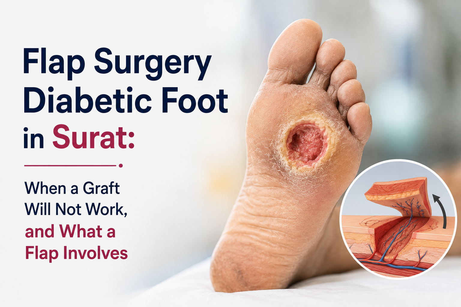
Flap surgery diabetic foot in Surat means moving healthy tissue, with its own blood supply, to cover a defect that cannot heal on its own. It is used when bone, tendon or joint is exposed, because a skin graft cannot survive on those surfaces.
Reconstructive surgeons work up a ladder, starting with the simplest option that will work and climbing only when it will not. Understanding where a flap sits on that ladder explains both why it is offered and why it is not offered sooner.
Rung 1: why not just dressings?
Dressings and negative pressure therapy are the first rung, and they close many wounds without surgery at all. They work when the wound is small, the blood supply is adequate, infection is controlled and pressure has been removed.
When you must climb higher: the wound has stalled for weeks despite proper care, it is too large to close by contraction, or bone or tendon is visible at the base. Wound care itself continues at every rung, managed under non-surgical wound management.
Rung 2: why not simply stitch it closed?
Direct closure is the next rung and the simplest surgical option. The wound edges are brought together and sutured, which works well for small defects where there is enough loose surrounding skin.
When you must climb higher: the defect is too wide, or pulling the edges together would put the closure under tension. Tension is the enemy here, because a tight closure on a diabetic foot cuts off its own blood supply and breaks down within days.
Rung 3: why not a skin graft?
A skin graft is a thin sheet of skin laid onto the wound, and it closes large areas efficiently. It is the workhorse of foot wound closure and is described fully in our post on skin grafting for diabetic wounds.
When you must climb higher: a graft has no blood supply of its own and survives only by growing new vessels from the surface beneath it. That means it needs a vascular bed. It will not take on exposed bone, bare tendon or an open joint, and it does poorly on weight-bearing surfaces like the heel where it stays thin and fragile.
This is the single most important point on this page. When someone says a graft was tried and failed on an area of exposed bone, the graft did not fail, it was never going to work.
Rung 4: what does a flap do differently?
A flap brings its own blood supply with it. Because the tissue arrives already perfused, it can survive over bone, tendon and joint, and it provides thicker, more durable padding for areas that take body weight.
- It covers structures a graft cannot, including exposed bone and tendon.
- It brings blood supply into the area, which also helps control infection.
- It provides durable padding for the heel and sole.
- It allows deeper reconstruction, including filling dead space left after debridement.
What types of flap are used on the foot?
Flaps are chosen by defect size, location and available blood supply. Local flaps move nearby tissue, regional flaps bring tissue from further up the leg, and free flaps transfer tissue from elsewhere in the body with the vessels rejoined under a microscope.
| Type | Where the tissue comes from | Typically used for |
|---|---|---|
| Local flap | Skin and tissue immediately beside the defect | Small to moderate defects with healthy surrounding tissue |
| Regional / pedicled flap | Further up the foot or leg, kept attached to its own vessels | Medium defects, ankle and heel cover |
| Free flap | A distant site such as the thigh or abdomen, vessels rejoined microsurgically | Large defects, poor local tissue, major limb salvage |
| Muscle flap | Local or distant muscle, often with a graft over it | Filling deep dead space and helping control infection |
What happens during flap surgery?
Flap surgery is done under general or regional anaesthesia and takes longer than a graft, particularly a free flap. The wound is fully debrided, the flap is raised with its blood vessels, moved into position and sutured, and for a free flap the vessels are joined under a microscope.
- Circulation assessment first, through our vascular services, because a flap needs vessels to connect to.
- Complete debridement of dead and infected tissue, including infected bone where present, as discussed in bone infection of the diabetic foot.
- Planning and raising the flap, preserving its vascular pedicle.
- Insetting the flap into the defect without tension.
- Microvascular anastomosis for a free flap, joining artery and vein under magnification.
- Donor site closure, directly or with a skin graft.
- Immobilisation and monitoring in a protected position.
Why are the first few days so closely watched?
Because a flap's blood supply can fail, and the first forty-eight to seventy-two hours are when it usually happens. The flap is checked frequently, often hourly at first, for colour, temperature, swelling and refill, since a failing flap can sometimes be rescued if returned to theatre quickly.
That is the reason for what can feel like excessive observation. A flap that turns pale or dusky is not left until the morning round; it goes back to theatre. Knowing this in advance makes the intensity of the first days easier to understand.
What are the risks?
Risks include partial or complete flap failure, infection, bleeding or clot at the vessel join, wound breakdown at the edges, donor site problems and the need for further surgery. In diabetes, healing is slower and infection risk is higher throughout.
- Vascular failure of the flap, partial or complete.
- Thrombosis at the anastomosis in free flaps.
- Infection in an already compromised foot.
- Edge necrosis or wound breakdown needing a further procedure.
- Donor site pain, scarring or delayed healing.
- Bulky flap initially, sometimes needing thinning later.
These are weighed against the alternative. For a defect with exposed bone that no graft can cover, the realistic comparison is not flap versus graft, it is flap versus amputation, discussed further in our post on limb preservation surgery.
What is recovery like?
Expect several days in hospital with the foot elevated and immobilised, then a graduated return to weight bearing over weeks. Most flaps are stable by two to three weeks, but full maturation of the tissue takes months.
- Days 1 to 3: strict elevation, immobilisation, frequent flap monitoring.
- Days 3 to 14: monitoring eases, dressings and donor site care, sutures reviewed.
- Weeks 2 to 6: graduated weight bearing as instructed, physiotherapy begins.
- Weeks 6 to 12: return to walking in protective footwear, flap softens.
- Months 3 to 12: flap matures and thins, custom insoles fitted, long term protection under preventive foot care.
Smoking materially worsens flap outcomes by constricting the small vessels the flap depends on. Stopping before surgery is one of the few things entirely within the patient's control.
When is a flap not possible?
A flap is not always the right answer. It needs vessels to connect to, a patient fit enough for longer surgery, and infection that can be controlled. Where those are missing, a different plan is safer.
- Severely blocked arteries that cannot be reopened.
- Uncontrolled spreading infection or sepsis, which must be treated first.
- General health that cannot tolerate long anaesthesia or staged surgery.
- Tissue loss so extensive that no useful weight-bearing foot would remain.
- Inability to comply with immobilisation, which the flap depends on.
In those situations the honest options are continued wound management, a smaller procedure, or a planned amputation. Being told a flap is not advisable is not the same as being abandoned; it usually means the risk of a failed flap outweighs its benefit.
Flap surgery and limb salvage at our Surat clinic
Elegance Diabetic Foot & Ulcer Clinic is based in Surat and led by Dr. Ashutosh Shah, a plastic and microvascular surgeon. For flap surgery diabetic foot in Surat, the clinic provides circulation assessment, debridement, local and free flap reconstruction, and the follow-up needed afterwards, with the specialty background explained in our post on choosing a reconstructive foot surgeon. The wider range of procedures is listed under diabetic foot surgery, and related problems under foot conditions we treat.
You can also send clear photos of the wound, along with any reports, to our team on WhatsApp for initial guidance and book a consultation. If there is fever, spreading redness or foul discharge, seek care the same day. Follow EDFC on Facebook, Instagram and YouTube for real limb salvage stories.
Climb only as high as you need to
The right operation is the simplest one that will actually work. If a wound has exposed bone or tendon, no amount of dressing or grafting will close it, and recognising that early is what prevents months of failed attempts on a foot that has time to lose.
Next step: book a wound and circulation assessment with Dr. Ashutosh Shah at Elegance Diabetic Foot & Ulcer Clinic, Surat, call +91 83205 00350, or send photos and reports on WhatsApp today.
Medical disclaimer: This article is for educational purposes only and is not a substitute for professional medical diagnosis or treatment. Whether a flap is possible depends on circulation, infection, tissue loss and general fitness, and can only be judged after in-person assessment. Fever, spreading redness or foul discharge needs urgent care. Please consult Dr. Ashutosh Shah or a qualified specialist about your condition. For further guidance, see the NHS guide to plastic surgery and the IWGDF diabetic foot guidelines.
quiz Frequently Asked Questions
This article is general education, not a diagnosis. If you have a diabetic foot wound, please have it assessed in person. Send a photo on WhatsApp or book a consultation.
About the Author
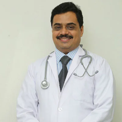
Dr. Ashutosh Shah, Plastic, Reconstructive & Diabetic Foot Surgeon, Elegance Diabetic Foot & Ulcer Clinic, Surat
Dr. Ashutosh Shah is a board certified plastic and reconstructive surgeon with over 22 years of experience and more than 10,000 limbs and feet saved. He founded Elegance Diabetic Foot & Ulcer Clinic, India's first chain of clinics dedicated to foot care, with centres in Surat and OPD partners across South Gujarat. He is known for a limb first approach, safe wound care and ethical, natural results.