
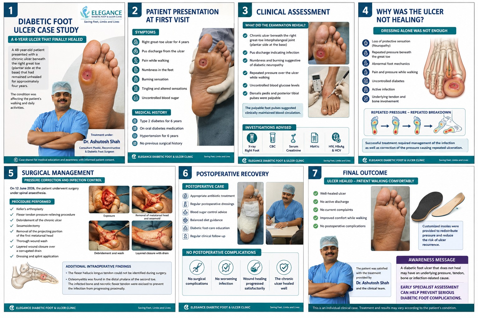
This diabetic foot ulcer case study follows a 48-year-old patient whose ulcer beneath the right great toe had remained unhealed for approximately four years. Assessment identified more than a surface wound: repeated pressure, diabetic neuropathy, infection and underlying structural problems had to be addressed before sustained healing could occur.
A Diabetic Foot Ulcer That Had Not Healed for Four Years
A 48-year-old patient presented to Elegance Diabetic Foot & Ulcer Clinic (EDFC) with a chronic ulcer beneath the right great toe, around the plantar side of the interphalangeal joint.
The ulcer had remained unhealed for approximately four years and was affecting the patient's walking and routine daily activities.
This was no longer simply a surface wound requiring another dressing. Its long duration raised an important clinical question: why was the ulcer repeatedly failing to heal?
The patient was assessed under the care of Dr. Ashutosh Shah, Consultant Plastic, Reconstructive & Diabetic Foot Surgeon, with the aim of identifying and treating the factors maintaining the ulcer.
What Did the Patient Have at the First Visit?
At presentation, the patient reported:
- Right great-toe ulcer for approximately four years
- Pus discharge from the ulcer
- Pain while walking
- Numbness in the feet
- Burning sensation
- Tingling and altered sensations
- Uncontrolled blood glucose
The medical history documented type 2 diabetes for six years, treatment with oral diabetes medicines and hypertension for six years.
These findings were important because a non healing diabetic foot ulcer cannot always be understood by looking at the wound surface alone.
The clinical team needed to assess sensation, infection, circulation, repeated pressure and the deeper structures beneath the ulcer.
Why Was This Diabetic Foot Ulcer Not Healing?
Several factors appeared to be contributing to the persistence of the ulcer.
A chronic wound needs more than repeated dressing when pressure, neuropathy, infection or deeper tissue involvement continues to interfere with healing. A structured diabetic foot ulcer treatment approach considers the wound together with the factors responsible for its persistence.
Loss of Protective Sensation
The patient's numbness, burning and altered sensation were suggestive of diabetic neuropathy.
Protective sensation normally warns a person about excessive pressure, friction, heat or injury. When this warning system becomes impaired, a patient may continue walking on an overloaded area without realising that tissue damage is occurring.
Repeated Pressure Beneath the Great Toe
The ulcer was located on a plantar, weight-bearing area beneath the great toe.
This meant that walking could repeatedly expose the wound to mechanical stress. Even when dressing temporarily improved the wound surface, continued loading could contribute to further tissue breakdown.
This is why pressure management is an important part of chronic diabetic foot ulcer treatment.
Abnormal Foot Mechanics
Abnormal mechanics can concentrate pressure over a relatively small part of the foot.
When abnormal loading occurs together with diabetic neuropathy, repeated tissue damage can happen without the level of pain that would normally make a person stop walking on the area.
Active Infection
The patient had pus discharge from the ulcer, indicating infection.
In a chronic diabetic foot wound, infection needs to be assessed for its depth and severity rather than simply covered with a dressing.
Uncontrolled Blood Glucose
Uncontrolled blood glucose was another important factor documented in this case.
Diabetes management therefore needed attention alongside local wound care, infection control and pressure management.
Why Was the Diabetic Foot Ulcer Not Healing Despite Regular Dressings
Dressings are important, but a dressing cannot necessarily remove the reason a wound keeps breaking down.
In this case, the clinical picture suggested a cycle such as:
Repeated pressure → tissue damage → ulcer → dressing → continued pressure → repeated breakdown
If the patient continues loading the same high-pressure point with every step, changing the dressing does not automatically remove the mechanical cause.
This is an important principle in diabetic foot ulcer healing.
For a chronic plantar ulcer, the clinical team needs to ask not only:
“What dressing should be applied?”
but also:
“Why does this exact area keep breaking down?”
What Did the Clinical Assessment Reveal?
Clinical examination documented a chronic ulcer beneath the right great-toe interphalangeal joint on the plantar side.
The assessment identified:
- Pus discharge indicating infection
- Numbness and burning suggestive of diabetic neuropathy
- Repeated pressure over the ulcer while walking
- Uncontrolled blood glucose
The dorsalis pedis and posterior tibial pulses were palpable. In this patient, the finding suggested clinically maintained circulation.
However, palpable pulses alone should not be treated as a complete vascular assessment when peripheral arterial disease is clinically suspected.
Which Investigations Were Advised?
The case documents the following investigations:
- X-ray of the right foot
- Complete blood count (CBC)
- Serum creatinine
- HbA1c
- HIV testing
- HBsAg
- HCV testing
The investigations required for a diabetic foot ulcer depend on the individual clinical findings.
Imaging can become particularly important when a clinician suspects deeper infection, structural abnormalities or possible bone involvement.
Can Bone Infection Hide Under a Diabetic Foot Ulcer?
Yes. In some patients, infection from a diabetic foot ulcer can extend into the underlying bone and cause osteomyelitis.
In this individual case, osteomyelitis and necrotic bone were identified during surgery.
When pus, increasing discharge or deeper infection is suspected, appropriate diabetic foot infection treatment depends on the depth and severity of infection, tissue involvement and whether bone infection is present.
Not every diabetic foot ulcer involves bone. The possibility of osteomyelitis needs to be assessed from the overall clinical picture, examination, imaging and other investigations when indicated.
This distinction matters because diabetic foot ulcer infection treatment can be very different when infection extends beyond superficial soft tissue.
How Was the 4-Year Non-Healing Diabetic Foot Ulcer Treated?
After assessment, the patient underwent surgery on 12 June 2026 under spinal anaesthesia.
The treatment was designed not only to manage the ulcer itself but also to address infection and pressure-related factors contributing to repeated breakdown.
The procedures documented in this individual case included:
- Keller's arthroplasty
- Flexor tendon pressure-relieving procedure
- Debridement of the chronic ulcer
- Sesamoidectomy
- Removal of the projecting portion of the first metatarsal head
- Thorough wound wash
- Layered wound closure over a corrugated drain
- Dressing and splint application
These procedures should not be interpreted as standard treatment for every chronic diabetic foot ulcer.
The need for surgery and the type of procedure used depends on ulcer location, infection, deformity, pressure distribution, tissue viability, bone involvement and the patient's overall health.
What Was Found During Surgery?
Surgery provided further information about the deeper condition of the foot.
According to the case documentation, the flexor hallucis longus tendon could not be identified during surgery.
Osteomyelitis was also found in the distal phalanx of the second toe. Infected bone and necrotic flexor tendon were excised to help prevent the infection from progressing proximally.
These findings highlight an important lesson from this diabetic foot ulcer case study: the appearance of a wound on the skin does not necessarily reveal everything occurring beneath it.
A small external opening can sometimes coexist with much deeper pathology.
Why Was Pressure Correction Important?
A plantar ulcer receives mechanical pressure when a person stands or walks.
If excessive pressure continues at the same location, wound healing can become difficult even when appropriate dressings are being used.
Effective diabetic foot offloading aims to reduce repeated mechanical stress at the ulcer site. Depending on the individual foot, this may involve specialized footwear, insoles, casts, removable devices or other pressure-relieving strategies.
In selected cases, surgery may be considered when a structural problem is responsible for persistent abnormal pressure.
In this patient, the surgical management included procedures intended to address the pressure contributing to recurrent tissue breakdown.
What Happened After Surgery?
Treatment did not end after the operation.
The postoperative plan documented in the case included:
- Appropriate antibiotic treatment
- Regular postoperative dressings
- Blood glucose control advice
- Balanced diet guidance
- Diabetic foot-care education
- Regular clinical follow-up
The case presentation reports no postoperative complications.
The wound subsequently progressed satisfactorily toward healing.
Final Outcome: The Chronic Ulcer Healed
At final follow-up, the ulcer was documented as well healed.
The patient had:
- No active discharge
- No current complaints
- Improved comfort while walking
- No documented postoperative complications
The final clinical photograph shows closure of the previously ulcerated plantar area.
For a patient who had lived with a chronic ulcer for approximately four years, the outcome was significant. Importantly, however, this is the result of one individual case and should not be used to predict the outcome of another patient's ulcer.
Why Were Customized Insoles Used After Healing?
Healing the ulcer did not mean that preventive care could stop.
The next objective was to reduce the chance of recurrence.
Customized insoles were provided in this case to help redistribute pressure and reduce excessive loading of vulnerable areas.
This is particularly important when neuropathy is present because a patient may not feel increasing pressure early enough to respond to it.
After diabetic foot ulcer healing, ongoing care may include:
- Appropriate footwear
- Pressure redistribution
- Daily foot inspection
- Blood glucose management
- Regular clinical follow-up
- Early treatment of callus or pressure areas
- Prompt assessment of new blisters or skin breakdown
A healed ulcer should therefore be considered a foot that still requires preventive attention.
What Can We Learn From This Diabetic Foot Ulcer Case Study?
The central lesson is not that every long-standing ulcer requires surgery.
Instead, this diabetic foot ulcer case study demonstrates why a persistent wound should trigger a search for the factors preventing healing.
Those factors may include:
Neuropathy + repeated pressure + abnormal mechanics + infection + poor glucose control + deeper tissue or bone involvement
Different patients may have different combinations of these problems.
Treating only the visible wound may leave the underlying cause unchanged. Effective management therefore starts with identifying why the ulcer is failing to heal.
In this patient, treating infection and addressing the pressure-related and structural factors formed part of the pathway to healing.
When Should a Non-Healing Diabetic Foot Ulcer Be Reassessed?
A diabetic foot ulcer should not continue receiving exactly the same treatment indefinitely when it is failing to make meaningful progress.
A comprehensive foot evaluation can assess the wound alongside sensation, circulation, pressure, deformity and other factors that may be delaying healing.
Reassessment may consider:
- Is pressure being adequately relieved?
- Is infection still present?
- Does the wound extend into deeper tissue?
- Is bone infection possible?
- Is circulation adequate for healing?
- Is glucose control interfering with recovery?
- Is footwear repeatedly loading the ulcer?
- Is an underlying deformity maintaining the pressure?
Increasing discharge, swelling, redness, foul smell, colour changes, fever or rapid wound deterioration require prompt medical assessment.
Conclusion
A non healing diabetic foot ulcer should not be viewed only as an open area of skin. When an ulcer persists despite repeated dressings, the reason for failed healing needs to be investigated.
This case demonstrates the importance of assessing neuropathy, repeated pressure, abnormal foot mechanics, infection, glucose control and possible deeper tissue or bone involvement.
At Elegance Diabetic Foot & Ulcer Clinic (EDFC), this patient was assessed and treated under Dr. Ashutosh Shah, Consultant Plastic, Reconstructive & Diabetic Foot Surgeon. The treatment plan addressed the clinical problems identified in this individual case, followed by postoperative wound care and pressure redistribution after healing.
The key educational message from this diabetic foot ulcer case study is simple: when a diabetic foot ulcer does not heal, identifying and treating the reason for non-healing is as important as caring for the wound itself.
quiz Frequently Asked Questions
This article is general education, not a diagnosis. If you have a diabetic foot wound, please have it assessed in person. Send a photo on WhatsApp or book a consultation.
About the Author

Dr. Ashutosh Shah, Plastic, Reconstructive & Diabetic Foot Surgeon, Elegance Diabetic Foot & Ulcer Clinic, Surat
Dr. Ashutosh Shah is a board certified plastic and reconstructive surgeon with over 22 years of experience and more than 10,000 limbs and feet saved. He founded Elegance Diabetic Foot & Ulcer Clinic, India's first chain of clinics dedicated to foot care, with centres in Surat and OPD partners across South Gujarat. He is known for a limb first approach, safe wound care and ethical, natural results.