Written by Dr. Ashutosh A Shah, M.B.B.S., M.S., M.Ch., D.N.B., Consultant Plastic and Reconstructive Surgeon, Elegance Diabetic Foot and Ulcer Clinic, Surat. Reg. no. [REG NO]. 22+ years in reconstructive surgery and limb salvage.
Medically reviewed by Dr. Ashutosh A Shah · Published [DD Month 2026] · Last reviewed [DD Month 2026]
A diabetic foot ulcer that has not improved in four weeks usually has one of four reasons: poor blood supply, infection including bone infection, continued pressure on the wound, or uncontrolled blood sugar. Advanced treatment means identifying which applies, then debridement, offloading, infection control and restoring blood flow as needed.
If you have been dressing a wound at home for weeks and it is the same size it was, that is not bad luck and it is not a matter of finding a better ointment. A wound that is not healing is a wound that is being stopped by something, and there is a short list of things that stop it.
This page explains that list, how each one is identified, and what is actually done about it. If you only have a minute, read section 10 first.
What counts as a non-healing ulcer, and when should you worry?
A wound that is healing gets visibly smaller. If yours has not, four weeks is the point at which you stop waiting.
The international guidelines on diabetes-related foot disease are specific about this. Where an ulcer fails to show signs of healing within four to six weeks despite optimal management, the recommendation is to consider angiography and revascularisation, which in plain terms means investigating whether blood is reaching the wound at all.
Note the phrase "despite optimal management". Four weeks of a dressing changed at home is not optimal management, so the clock in practice starts when proper treatment starts. If nothing proper has started yet, that is the thing to fix this week.
How to judge your own wound honestly. Photograph it in the same light, from the same distance, with a ruler or a coin beside it, once a week. Memory is unreliable and daily change is invisible. A photograph from three weeks ago is not.
What "improving" looks like: the edges are pulling inwards, the base is pink or red rather than yellow or black, discharge is reducing, and the surrounding skin is calm. If those are not happening, the wound is stalled even if it is not obviously worse.
What are the four reasons an ulcer stops healing?
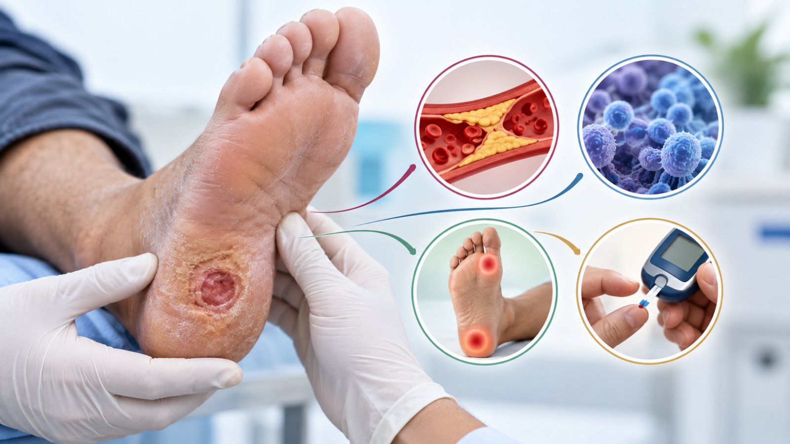
Almost every stalled diabetic foot ulcer is stopped by one or more of four things. The whole purpose of an assessment is to work out which.
| Cause | How it is identified | What it needs |
|---|---|---|
| Poor blood supply | Feeling for pulses in the foot, Doppler assessment of the waveform, ankle brachial index and toe brachial index | Assessment by a vascular team, and where indicated angiography and restoring the blood flow |
| Infection, including bone infection | Clinical signs, the probe-to-bone test, and imaging where bone involvement is suspected | Appropriate antibiotics directed by culture, drainage where there is pus, and surgery where bone is involved |
| Continued pressure on the wound | Site of the ulcer, the pattern of callus around it, and how you walk | Offloading, ideally with a device you cannot take off |
| Uncontrolled blood sugar | Blood glucose and HbA1c, reviewed with your treating physician | Optimising control, with insulin if necessary |
Two things follow from that table and they are worth stating plainly.
You cannot tell which one applies by looking. A wound stopped by poor blood supply and a wound stopped by pressure can look identical. That is why assessment comes before treatment, and why a dressing chosen without an assessment is a guess.
More than one is usually present. Treating the infection while the pressure continues, or offloading beautifully while blood is not reaching the foot, gets you nowhere. All the active causes have to be addressed together.
The guidelines also list what else should be attended to alongside the wound: swelling, nutrition, cardiovascular risk factors and the psychological strain of a long illness. That last one is real. Months of dressings and restricted walking wear people down, and a patient who has given up is a patient whose wound will not heal.
Why can a wound not heal without blood supply?
Because healing is an active, energy-hungry process, and everything it needs arrives through the arteries.
Oxygen, white cells to fight infection, the building blocks for new tissue and antibiotics all reach a wound through blood. If the arteries supplying the foot are narrowed, none of them arrive in sufficient quantity, and no dressing, ointment or advanced therapy compensates for that. This is why blood supply is assessed first.
How it is assessed. The guidelines describe feeling the pulses in the foot, assessing the Doppler waveform, and measuring the ankle brachial index and toe brachial index. Peripheral arterial disease is less likely where the pedal Doppler waveform is triphasic or biphasic, the ABI is between 0.9 and 1.3, and the TBI is 0.70 or above. These are quick, painless bedside tests.
What happens if the blood supply is poor. Angiography and restoring the flow are considered. The guidelines flag increased urgency where the ankle pressure is below 50 mmHg or the ABI is below 0.4.
Vascular assessment and revascularisation are the territory of vascular surgery and interventional radiology, so this part of your care is arranged with them rather than done by any single clinic alone. Any service that tells you it can heal an ischaemic foot without addressing the circulation is describing something that is not possible.
How is bone infection different from skin infection?
It is deeper, it is harder to clear, and it changes the whole plan. Distinguishing the two is one of the most important steps in the assessment.
How infection is diagnosed. The guidelines require at least two clinical signs or symptoms of inflammation, meaning redness, warmth, induration, and pain or tenderness, or else purulent secretions. Note that infection is a clinical diagnosis, made by examining the foot, rather than something a swab decides on its own.
The probe-to-bone test. This is the simple, decisive bedside test, and it is worth knowing the name so you can ask whether it has been done. A sterile metal probe is passed gently into the ulcer to determine whether bone can be touched or seen. A positive result, alongside an abnormality on imaging, points towards osteomyelitis, meaning infection of the bone.
Why the distinction matters so much:
- Skin and soft tissue infection is usually treated with a course of antibiotics chosen by culture, plus drainage if there is pus
- Bone infection needs a longer course, and often surgery to remove the infected bone, because antibiotics alone frequently do not clear it
- A wound sitting over infected bone will not close however well it is dressed, which is one of the commonest reasons an ulcer runs for months
If your wound has been treated as a surface problem for weeks with no progress, ask directly whether the probe-to-bone test has been done and whether imaging has been considered.
Why is offloading the step most often skipped?
Because it is the one that inconveniences the patient, and because it works only if it is actually worn.
Most diabetic foot ulcers sit on the sole and are caused by pressure. Every step reapplies the force that created the wound. A wound under repeated pressure cannot close, however good the dressing.
The guideline position is unambiguous: the preferred offloading treatment for a neuropathic ulcer on the sole is a non-removable knee-high device, meaning a total contact cast or a walker rendered irremovable. Non-removable, because a device that can be taken off is taken off, usually for a few steps to the bathroom, and those steps are enough.
That is not a comment on anyone's discipline. It is a recognised clinical reality, and it is the reason the guideline specifies non-removable rather than simply recommending a boot.
The devices themselves, how they differ, how long each is worn and what daily life in one is actually like are covered on our separate page on offloading footwear and aircast boots. For this page the point is simply that if nobody has addressed pressure, your wound has not yet had proper treatment.
What is debridement, and why is it repeated?
Debridement is the removal of dead tissue and callus from the wound so that living tissue can heal. The guidelines describe sharp surgical debridement as the standard method.
Why it is necessary. Dead tissue is a physical barrier and a home for bacteria. A wound cannot close over it. The thick callus that builds around a pressure ulcer also concentrates force onto the wound edges, making things worse.
Why it is repeated. Dead tissue and callus re-form. Debridement is usually a series of small procedures over weeks rather than one event, and needing it again is not a sign of failure.
What it is not. It is not something to attempt at home. Cutting callus yourself, particularly with reduced sensation, is one of the commonest ways people turn a small problem into a large one.
Does it hurt? Frequently less than expected, because many people with diabetic foot ulcers have reduced sensation in the area. Where sensation is intact, local anaesthetic is used. Tell the clinician what you feel rather than enduring it, because pain during debridement is also clinical information.
One important note on method. The guidelines make strong recommendations against autolytic, biosurgical, hydrosurgical, chemical, laser and ultrasonic debridement in place of standard sharp debridement. If something other than sharp debridement is being proposed, it is fair to ask why.
What advanced wound therapies exist, and when is each appropriate?
Several exist. All of them are adjuncts, all of them come after standard care rather than instead of it, and the evidence behind them is weaker than the marketing around them.
Standard care means sharp debridement, offloading, dressings that keep the wound appropriately moist, and management of infection. The guideline on wound healing interventions is explicit that its recommendations apply only once best standard care has failed to heal the ulcer.
What it supports, all as conditional recommendations:
| Therapy | When it is considered | Strength of the recommendation |
|---|---|---|
| Sucrose-octasulfate impregnated dressing | Neuro-ischaemic ulcers, after two weeks of unsuccessful standard care | Conditional, moderate certainty |
| Autologous leucocyte, platelet and fibrin patch | Hard-to-heal ulcers, where the resource exists | Conditional, moderate certainty |
| Hyperbaric oxygen | Neuro-ischaemic or ischaemic ulcers, as an adjunct | Conditional, low certainty |
| Topical oxygen | Wounds not responding to standard care | Conditional, low certainty |
| Placental-derived products | Where standard care has failed | Conditional, low certainty |
| Negative pressure wound therapy | Post-surgical wounds only. See below | Conditional, low certainty |
The negative pressure point deserves emphasis, because clinics blur it. The guideline supports NPWT conditionally for post-surgical wounds, and makes a strong recommendation against using it for non-surgically related diabetic foot ulcers. Same machine, opposite recommendations, depending entirely on how the wound came about. If NPWT is being proposed for you, ask which of those two situations you are in.
The guideline also suggests against the routine use of cellular and acellular skin substitutes, other cell therapies and growth factor therapy. And it says of its own evidence base that the certainty of much of it remains poor overall.
Read that as useful rather than discouraging. It means the boring things, debridement, offloading, infection control and blood supply, are where the results come from, and that anyone selling you an advanced therapy before those four are sorted has the order wrong.
What should you never put on a diabetic foot ulcer?
This section exists because the wrong thing is applied to diabetic foot wounds in Indian homes every day, usually by someone trying to help.
The guidelines make strong recommendations against a list of things, and strong is the highest strength they use. Among them:
- Honey and bee-related products. A strong recommendation against, despite the reputation.
- Any dressing or topical application impregnated with herbal remedies. This includes turmeric pastes, herbal oils and traditional preparations applied to the wound.
- Topical antiseptic or antimicrobial dressings used for wound healing. Household antiseptics poured onto a wound are not treatment and can damage the healing tissue.
- Collagen or alginate dressings.
- Topical phenytoin.
- Other gases, including cold atmospheric plasma, ozone, nitric oxide and carbon dioxide.
- Physical therapies, including ultrasound, electrical stimulation and laser therapy for wound healing.
- Vitamin and trace element supplements, and agents that stimulate red cell production or supplement protein, taken for the purpose of wound healing.
Why this matters more than it sounds. A wound with something inappropriate on it is a wound whose appearance is altered, whose progress cannot be judged accurately, and which may be irritated by what has been applied. It also means weeks pass. The harm is rarely the substance itself. The harm is the delay.
The other rule. Do not cut, file or pare callus at home, and do not apply heat. Reduced sensation means you will not feel the damage while you do it.
If you have already been applying something, do not feel accused, and do say so at the appointment. Knowing what has been on the wound genuinely helps the person assessing it. Our home care guidance sets out what is appropriate to do between visits.
When does an ulcer need surgery?
Surgery here usually means one of four things, and none of them automatically means amputation.
- Debridement in theatre, where the dead tissue is extensive or the wound needs exploring properly under anaesthesia.
- Drainage, where there is a collection of pus. This is often urgent.
- Removal of infected bone, where osteomyelitis has been confirmed and antibiotics alone will not clear it.
- Reconstruction and soft tissue cover, using grafts or flaps to close a defect that will not close on its own. This is the limb salvage end of the work.
Surgery to restore blood flow sits alongside these and is arranged with the vascular team.
Our approach to limb salvage is described on the programs page. The broader question of how amputation risk is reduced over time is covered separately on our page about preventing diabetic foot amputation, which is the right page to read if that is the fear behind your search.
Which signs mean go today, not next week?
These are the same-day signs. If any of them applies, this is not a wait-and-see situation.
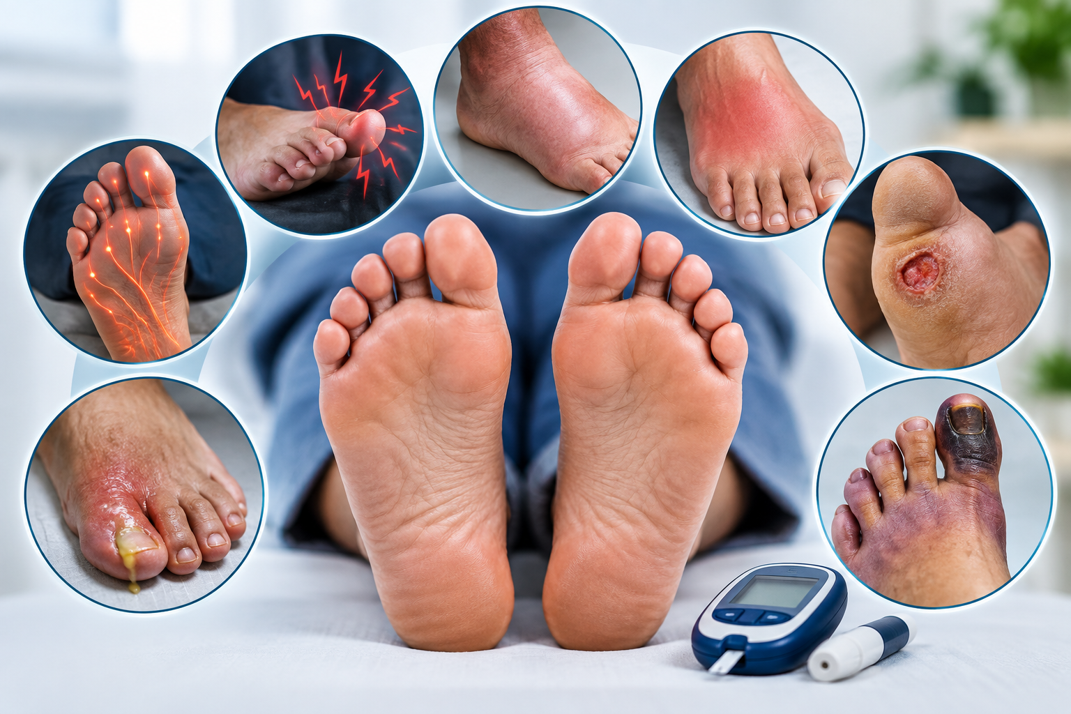
- Redness spreading outwards from the wound, particularly if it is advancing up the foot or leg
- Fever, chills, or feeling generally unwell alongside a foot wound
- A foul smell from the wound or the dressing
- Black tissue appearing anywhere on the foot or toes
- Sudden severe pain in a foot that was previously not very painful
- A wound that suddenly stops hurting. This one is missed constantly because it feels like improvement. It can mean nerve damage has progressed or tissue has died, and it is a reason to be seen rather than relieved
Also go the same day for: pus discharging, a rapid increase in swelling, blood sugar suddenly running much higher than usual with no other explanation, or a wound that has visibly deepened in a day or two.
If you are reading this on behalf of a parent, the most useful thing you can do tonight is look at the foot yourself rather than take their word for it. People with reduced sensation genuinely underestimate what is happening, because it does not hurt.
What happens at an assessment at EDFC, Surat
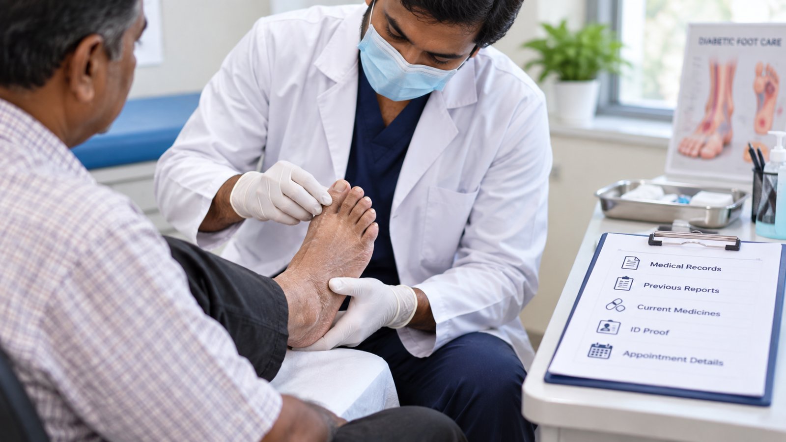
The assessment exists to answer one question: which of the four causes is stopping your wound, and in what combination.
What it involves in practice: examination of the wound and the whole foot, assessment of the circulation including pulses, Doppler and pressure indices, testing for loss of protective sensation with a monofilament or tuning fork, the probe-to-bone test where bone involvement is a possibility, culture where infection is suspected, imaging where indicated, and a review of your blood sugar control with your treating physician.
From that comes a plan that names the cause, sets out the treatment in order, and gives a review date at which progress is measured rather than estimated.
Dr. Ashutosh A Shah is a Consultant Plastic and Reconstructive Surgeon with M.Ch. and D.N.B. qualifications and over 22 years in reconstructive practice, which covers the debridement, soft tissue cover and reconstruction side of limb salvage. Vascular assessment and revascularisation are arranged with the vascular team, and diabetes control with your physician, because good diabetic foot care is a team activity rather than a single specialty.
Three things you will not be told here. That a dressing alone will fix a stalled wound. That an advanced therapy should be tried before the basics are in place. Or a healing time before anyone has worked out what is stopping it.
Next step
If a wound has not changed in four weeks, the useful next step is an assessment that names the cause, not another dressing.
- Photograph the wound today with a ruler or coin beside it, so progress can be measured later
- Write down when it started and what has been applied to it, including anything from home
- Bring your recent blood sugar and HbA1c results, and a list of your medicines
- Bring the footwear you actually wear every day, not your best pair
Book a diabetic foot assessment at EDFC, Surat
Medical disclaimer. This article is general health information and is not a substitute for examination, diagnosis or treatment by a qualified doctor. It does not describe anything that should be attempted at home. Do not debride, cut or pare a wound or callus yourself, and do not apply any substance to a diabetic foot ulcer without medical advice. Seek medical care the same day for spreading redness, fever, foul smell, black tissue, pus, sudden severe pain, rapidly increasing swelling, or a wound that suddenly stops hurting.
quiz Frequently Asked Questions
This article is general education, not a diagnosis. If you have a diabetic foot wound, please have it assessed in person. Send a photo on WhatsApp or book a consultation.
About the Author

Dr. Ashutosh Shah, Plastic, Reconstructive & Diabetic Foot Surgeon, Elegance Diabetic Foot & Ulcer Clinic, Surat
Dr. Ashutosh Shah is a board certified plastic and reconstructive surgeon with over 22 years of experience and more than 10,000 limbs and feet saved. He founded Elegance Diabetic Foot & Ulcer Clinic, India's first chain of clinics dedicated to foot care, with centres in Surat and OPD partners across South Gujarat. He is known for a limb first approach, safe wound care and ethical, natural results.