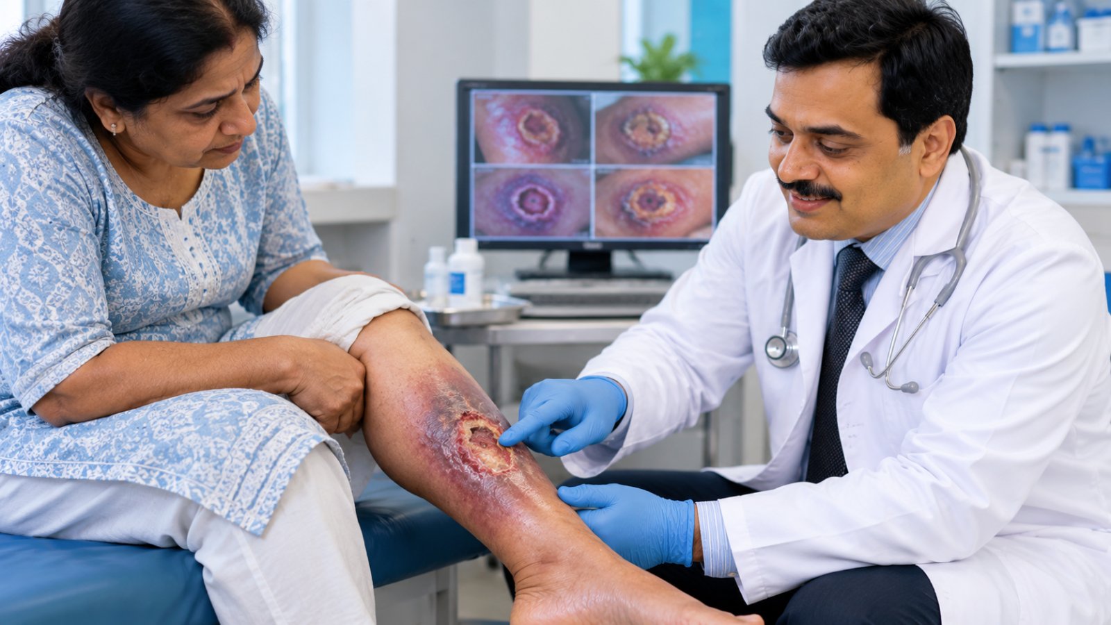
Pyoderma gangrenosum is an inflammatory, non-infectious ulcer that starts as a painful pustule and grows fast with a purple, undermined edge. It is often mistaken for infection and debrided, but cutting triggers pathergy and the ulcer enlarges. Diagnosis rests on clinical criteria and a biopsy to exclude other causes; treatment is immune-calming medicine, not surgery.
A leg ulcer that keeps growing despite antibiotics and wound cleaning can be confusing for patients and doctors.
Most chronic ulcers develop because of poor circulation, diabetes, pressure or infection. However, some ulcers result from an abnormal inflammatory response.
A pyoderma gangrenosum leg ulcer may initially look like an ordinary infected wound. Its appearance and rapid progression can lead to repeated antibiotic courses or surgical procedures that fail to improve the condition.
Understanding the warning signs helps patients seek appropriate specialist assessment.
Key Takeaways
-
Pyoderma gangrenosum is a rare inflammatory skin disease, not primarily a bacterial infection.
-
It often begins as a painful pustule, bump or small area of skin breakdown.
-
The ulcer may enlarge rapidly and develop a violet or purple undermined border.
-
Pain is often severe compared with the visible wound.
-
Surgical trauma can trigger pathergy and worsen the ulcer in susceptible patients.
-
Diagnosis relies on clinical findings, examination and tests to exclude other causes.
-
Treatment commonly involves anti-inflammatory or immune-modulating medicines.
-
Dermatology and wound-care specialists may need to work together.
What Is Pyoderma Gangrenosum?
Pyoderma gangrenosum is an uncommon inflammatory condition belonging to a group of disorders called neutrophilic dermatoses.
Neutrophils are white blood cells that normally help the body respond to infection.
In pyoderma gangrenosum, abnormal inflammatory activity contributes to tissue damage and ulcer formation.
Despite its name, the condition is not caused by pus-producing bacteria and is not the same as gangrene from inadequate blood supply.
How Does a Pyoderma Gangrenosum Ulcer Begin?
The condition may start as:
-
A small painful red bump
-
A pustule
-
A blister
-
A tender area of skin breakdown
The lesion may then develop into an ulcer.
Some ulcers enlarge quickly over days or weeks.
What Does the Ulcer Look Like?
Typical findings can include:
-
Severe pain
-
A rapidly enlarging wound
-
Violet or purple wound edges
-
Undermined borders, where tissue beneath the edge is damaged
-
Irregular ulcer shape
-
Surrounding redness or inflammation
The appearance varies between patients.
Not every painful ulcer with a purple edge is pyoderma gangrenosum.
Read more about Pyoderma gangrenosum and its clinical presentation.
How Is Pyoderma Gangrenosum Different From an Infected or Diabetic Leg Ulcer?
Distinguishing these conditions is important because their treatments can differ substantially.
An infected leg ulcer may require antibiotics, drainage or surgical removal of infected tissue.
A diabetic neuropathic ulcer often requires pressure relief, callus management and appropriate wound care.
Pyoderma gangrenosum generally requires treatment aimed at controlling inflammation.
Pyoderma Gangrenosum vs Infected Leg Ulcer vs Diabetic Ulcer
| Feature | Pyoderma Gangrenosum | Infected Leg Ulcer | Diabetic Neuropathic Ulcer |
|---|---|---|---|
| Pain | Often severe | Variable, frequently painful | May be painless due to neuropathy |
| Ulcer edge | May be violet and undermined | Often inflamed or irregular | Frequently surrounded by callus |
| Speed of growth | Can enlarge rapidly | May worsen as infection spreads | Often progresses with repeated pressure |
| Response to debridement | May worsen through pathergy | May require debridement when indicated | Often benefits from appropriate callus removal and debridement |
| Wound culture | May be negative or show colonisation or secondary infection | May identify pathogens | May show colonisation or infection |
| Associated conditions | Inflammatory bowel disease, arthritis, blood disorders | Infection and underlying wound causes | Diabetes, neuropathy and mechanical pressure |
| Main treatment approach | Control inflammation and protect the wound | Treat infection and underlying cause | Offloading, wound care and management of complications |
These features are clues rather than definitive diagnostic rules.
Can Pyoderma Gangrenosum Be Mistaken for Infection?
Yes.
The ulcer may look red, swollen and inflamed.
It may also produce discharge.
These findings can resemble a bacterial infection.
However, a positive wound culture does not automatically prove that infection is the primary cause.
Some wounds contain colonising bacteria without an invasive infection.
Pyoderma gangrenosum can also develop a genuine secondary infection.
Clinicians must assess both possibilities rather than assuming that they are mutually exclusive.
Why Can Surgical Debridement Make Pyoderma Gangrenosum Worse?
Debridement is the removal of dead, damaged or infected tissue.
It is an important treatment for many chronic wounds.
However, aggressive debridement of active pyoderma gangrenosum can sometimes cause the ulcer to enlarge.
The reason is a phenomenon called pathergy.
What Is Pathergy?
Pathergy is an exaggerated inflammatory response to relatively minor skin trauma.
In susceptible patients, an injury may trigger new inflammation or worsen an existing lesion.
Possible triggers include:
-
Surgical incisions
-
Sharp debridement
-
Needle punctures
-
Friction
-
Accidental skin injuries
Why Does the Ulcer Grow After Debridement?
During active pyoderma gangrenosum, cutting or scraping the wound may trigger additional inflammatory tissue damage.
The ulcer may then become larger or more painful.
This can create a difficult cycle:
-
The ulcer is mistaken for an infected wound.
-
Surgical debridement is performed.
-
The wound enlarges through pathergy.
-
The enlargement is interpreted as worsening infection.
-
Further debridement causes additional injury.
An ulcer getting bigger after debridement should prompt reconsideration of the diagnosis, particularly when pain is severe or the wound has characteristic inflammatory borders.
Does This Mean Surgery Is Always Wrong?
No.
Surgery is not automatically prohibited in every patient with pyoderma gangrenosum.
Urgent procedures may be necessary when another serious condition is present, such as a deep infection requiring source control.
Selected reconstructive procedures may also be considered after inflammatory activity is controlled.
The important point is that repeated aggressive debridement should not continue automatically when the clinical findings suggest pyoderma gangrenosum.
What Causes Pyoderma Gangrenosum?
The exact cause is not fully understood.
The condition involves abnormal immune activity and inflammation.
It is generally considered an autoinflammatory or immune-mediated skin disease rather than a conventional infection.
Which Diseases Are Associated With It?
Pyoderma gangrenosum may occur alongside:
Inflammatory bowel disease: Crohn's disease and ulcerative colitis.
Inflammatory arthritis: Including rheumatoid arthritis and certain other inflammatory joint conditions.
Blood disorders: Some haematological diseases, including selected blood cancers and monoclonal gammopathies.
Other inflammatory conditions: Less commonly, pyoderma gangrenosum may be associated with additional systemic disorders.
However, some patients have no identifiable associated disease.
Is Pyoderma Gangrenosum an Autoimmune Leg Ulcer?
The term autoimmune leg ulcer is sometimes used broadly, but pyoderma gangrenosum is more accurately described as a neutrophilic inflammatory disorder.
It is not identical to vasculitic ulcers, in which blood vessel inflammation can contribute to tissue damage.
Patients with unexplained inflammatory ulcers may need evaluation for several possible conditions.
Read about Autoimmune ulcers and their assessment.
How Is Pyoderma Gangrenosum Diagnosed?
There is no single blood test that confirms every case of pyoderma gangrenosum.
Diagnosis combines clinical findings, medical history, examination, biopsy and investigations for alternative causes.
1. Clinical Examination
The clinician assesses:
-
Ulcer appearance
-
Severity of pain
-
Speed of progression
-
Previous wound treatments
-
Possible pathergy
-
Associated medical conditions
2. Skin Biopsy
A biopsy is often used to help support the diagnosis and exclude other conditions.
The 2018 Delphi consensus diagnostic criteria include a biopsy from the ulcer edge demonstrating neutrophilic infiltration as the major criterion.
However, biopsy findings are not always specific, and a negative or inconclusive result does not necessarily exclude pyoderma gangrenosum.
Biopsy itself may trigger pathergy, so clinicians should plan sampling carefully.
3. Tests for Infection
Depending on the clinical findings, investigations may include:
-
Appropriate wound cultures
-
Tissue sampling
-
Blood tests
-
Imaging when deeper infection is suspected
These tests help distinguish primary infection from inflammation and identify secondary infection when present.
4. Circulation Assessment
Poor arterial blood flow can cause painful ulcers and tissue loss.
Clinicians may assess pulses and perform vascular investigations when indicated.
5. Excluding Other Causes
Important alternative diagnoses include:
-
Venous leg ulcers
-
Arterial ulcers
-
Diabetic ulcers
-
Vasculitis
-
Certain infections
-
Skin malignancies
-
Other inflammatory disorders
Patients with persistent diagnostic uncertainty may require assessment for Chronic ulcers of unknown cause.
What Are the Delphi Criteria and PARACELSUS Score?
The 2018 Delphi consensus criteria provide a structured approach using a major histological criterion and several minor clinical criteria.
The PARACELSUS score, described in 2019, is another diagnostic tool that considers findings such as rapid progression, exclusion of other causes, characteristic ulcer borders and severe pain.
These tools support clinical reasoning.
They should not replace specialist assessment or appropriate exclusion of other diagnoses.
What Treatments Help Control Pyoderma Gangrenosum?
Treatment depends on disease severity, ulcer size, pain, associated illnesses and infection risk.
The main objective is to control inflammation while protecting the wound.
1. Corticosteroids
Corticosteroids can reduce inflammatory activity.
Doctors may use topical, intralesional or systemic treatment depending on the condition.
Systemic corticosteroids require monitoring because they can increase blood glucose and infection risk.
This is especially important for patients with diabetes.
2. Ciclosporin
Ciclosporin is an immune-modulating medicine used in selected patients.
It can help control inflammatory activity but requires monitoring of kidney function, blood pressure and other potential adverse effects.
3. Biologic Medicines
Selected patients with difficult-to-control disease may receive biologic treatments.
Some therapies target inflammatory pathways, including tumour necrosis factor.
These treatments require specialist supervision and screening for relevant infections.
4. Gentle Wound Care
Wound care aims to:
-
Protect surrounding skin
-
Manage discharge
-
Reduce dressing-related trauma
-
Maintain a suitable wound environment
-
Address pain
-
Monitor for secondary infection
Non-adherent dressings may help reduce trauma during changes.
Read more about Wound care.
5. Treating Associated Conditions
If a patient has inflammatory bowel disease, arthritis or another associated condition, controlling that illness may be an important part of management.
Dermatology, rheumatology, gastroenterology and wound-care specialists may collaborate when necessary.
Are Antibiotics Useful?
Antibiotics do not treat the underlying inflammatory process of pyoderma gangrenosum.
However, they may be necessary if a genuine secondary bacterial infection is present.
Antibiotics should be prescribed according to clinical findings rather than ulcer appearance alone.
How Should the Ulcer Be Monitored During Healing?
Pyoderma gangrenosum often requires ongoing assessment because inflammatory activity and healing can change over time.
What Should Clinicians Monitor?
Useful findings include:
-
Ulcer dimensions
-
Pain intensity
-
Border inflammation
-
New lesions
-
Wound discharge
-
Surrounding skin condition
-
Treatment side effects
-
Signs of secondary infection
Can Wound Photographs Help?
Clinical photographs can document changes in wound size and appearance.
They may help clinicians compare progress between visits.
However, photographs should supplement rather than replace direct examination.
The frequency of monitoring depends on disease activity and treatment.
What Are Signs of Improvement?
Improvement may include:
-
Reduced pain
-
Less inflammation around the ulcer
-
Slower or stopped expansion
-
Development of healthy healing tissue
-
Gradual reduction in wound size
Complete healing may take time.
Some patients experience recurrence after improvement.
When Is Urgent Medical Care Needed?
Seek prompt assessment for:
-
Rapidly worsening pain
-
Fever or systemic illness
-
Spreading redness
-
Sudden wound enlargement
-
New black tissue
-
Significant bleeding
-
New circulation problems
These findings may indicate infection, vascular complications or other serious conditions.
Non Healing Leg Ulcer Specialist in Surat: When to Seek Assessment
Patients searching for a non healing leg ulcer specialist in surat should seek further evaluation when an ulcer continues to enlarge despite appropriate treatment, particularly if it is unusually painful or has a purple undermined border.
At Elegance Diabetic Foot & Ulcer Clinic (EDFC), Dr. Ashutosh A Shah, MBBS, MS, MCh, DNB, a Plastic and Reconstructive Surgeon, is associated with the assessment and management of complex wounds.
When pyoderma gangrenosum is suspected, collaboration with a dermatologist or rheumatologist may be appropriate.
The evaluation may include reviewing previous wound treatments, examining ulcer characteristics and arranging investigations to exclude infection, vascular disease and other causes.
Treatment decisions should follow the clinical diagnosis rather than assuming that every enlarging ulcer requires further surgery.
Conclusion
A pyoderma gangrenosum leg ulcer can resemble an infected wound, but its underlying cause and treatment may be very different.
Severe pain, rapid enlargement, purple undermined borders and worsening after debridement are important warning signs.
Aggressive surgical treatment during active disease can sometimes trigger pathergy and increase tissue damage.
Diagnosis requires careful clinical assessment, investigation and exclusion of alternative causes.
Treatment generally focuses on controlling inflammation, protecting the wound and managing associated conditions.
Early recognition can help prevent inappropriate repeated procedures and support safer wound management.
quiz Frequently Asked Questions
This article is general education, not a diagnosis. If you have a diabetic foot wound, please have it assessed in person. Send a photo on WhatsApp or book a consultation.
About the Author

Dr. Ashutosh Shah, Plastic, Reconstructive & Diabetic Foot Surgeon, Elegance Diabetic Foot & Ulcer Clinic, Surat
Dr. Ashutosh Shah is a board certified plastic and reconstructive surgeon with over 22 years of experience and more than 10,000 limbs and feet saved. He founded Elegance Diabetic Foot & Ulcer Clinic, India's first chain of clinics dedicated to foot care, with centres in Surat and OPD partners across South Gujarat. He is known for a limb first approach, safe wound care and ethical, natural results.