A leg ulcer unhealed after 6 weeks needs a structured workup, not a new dressing: an ankle-brachial pressure index, a venous duplex scan, sugar and kidney tests, a swab only if infected, and a biopsy if it looks unusual or has not improved after 12 weeks of correct care. The cause decides whether compression is safe.
A chronic leg ulcer can develop because of poor venous circulation, reduced arterial blood supply, diabetes-related complications or inflammatory diseases. Each cause requires a different treatment approach.
The most important step is identifying why the wound is not healing before choosing compression bandages, advanced dressings or other procedures.
Key Takeaways
-
A leg ulcer open for more than six weeks warrants a structured assessment of its underlying cause.
-
Venous, arterial, diabetic and vasculitic ulcers require different treatments.
-
ABPI helps assess arterial circulation before compression therapy.
-
Venous duplex ultrasound identifies venous reflux or obstruction.
-
HbA1c and kidney function tests help identify conditions affecting healing.
-
Wound swabs are generally reserved for suspected clinical infection, rather than routine sampling.
-
Unusual ulcers may require an early biopsy to investigate vasculitis, malignancy or other causes.
-
A wound that has not improved after approximately 12 weeks of appropriate care needs reassessment, including consideration of biopsy.
-
Compression can support venous ulcer healing, but it may be harmful when significant arterial disease is present.
When Does a Leg Ulcer Count as Chronic?
A leg ulcer is an open wound on the lower limb that does not heal normally.
Many clinical definitions describe a chronic wound as one that fails to progress through expected healing stages over several weeks.
For practical assessment, a wound remaining open for more than six weeks should raise concern about an underlying problem.
However, patients should not wait six weeks before seeking care. An ulcer with infection, severe pain, poor circulation or suspicious features requires earlier evaluation.
Why Do Some Leg Ulcers Remain Open?
Normal wound healing depends on adequate blood flow, healthy tissue, controlled inflammation and protection from repeated injury.
Healing may be delayed by:
-
Chronic venous insufficiency
-
Peripheral arterial disease
-
Diabetes and peripheral neuropathy
-
Infection
-
Kidney disease
-
Repeated pressure or trauma
-
Autoimmune disorders
-
Inflammatory skin diseases
-
Malignancy
-
Poor nutritional status
A leg wound not healing despite regular dressings may indicate that one or more of these factors has not been addressed.
Why Are Dressings Alone Sometimes Not Enough?
Dressings help protect a wound, manage moisture and support healing.
However, they cannot independently correct blocked arteries, venous reflux or systemic inflammatory disease.
For example, a venous ulcer may require compression, while an arterial ulcer may require urgent vascular assessment.
Using the same treatment for both conditions can delay healing or cause harm.
Which Tests Form the Basic Chronic Leg Ulcer Workup?
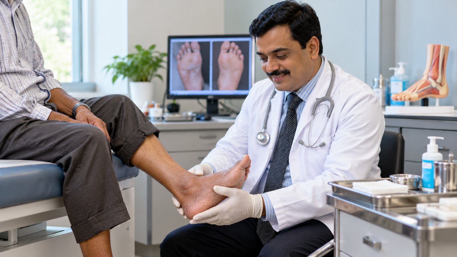
A proper assessment begins with medical history, examination of the wound and evaluation of circulation.
The clinician should document the ulcer's location, size, depth, appearance, surrounding skin and symptoms.
Tests are then selected according to the suspected cause.
Diagnostic Workup Table
| Test | What It Helps Identify | When It Is Done | Who May Order It |
|---|---|---|---|
| ABPI | Peripheral arterial disease and compression safety | Initial assessment when arterial disease must be excluded | Wound specialist or vascular clinician |
| Venous duplex ultrasound | Venous reflux, obstruction or thrombosis | Suspected venous disease | Vascular specialist |
| HbA1c | Diabetes or long-term glucose control | Initial assessment when indicated | Treating physician |
| Creatinine and eGFR | Kidney dysfunction affecting treatment and healing | Initial assessment when indicated | Treating physician |
| ESR or CRP | Inflammation or possible infection | When clinical findings suggest inflammation or infection | Treating physician |
| Autoimmune blood tests | Possible systemic autoimmune disease | When vasculitis or another inflammatory disorder is suspected | Appropriate specialist |
| Wound swab or tissue culture | Organisms associated with clinically suspected infection | When infection is suspected and sampling will guide treatment | Treating clinician |
| Skin biopsy | Vasculitis, malignancy or other atypical pathology | Suspicious ulcers or poor response to appropriate care | Dermatologist or appropriate specialist |
Not every patient requires every test.
The purpose is to use a logical diagnostic sequence rather than ordering investigations without a clinical indication.
Step 1: Examine the Wound
The clinician assesses:
-
Ulcer location
-
Wound dimensions
-
Tissue appearance
-
Surrounding skin changes
-
Pain severity
-
Signs of infection
-
Foot pulses
-
Sensation
-
Swelling
-
Previous wound history
The wound's location can provide useful clues, although location alone cannot establish the diagnosis.
Step 2: Assess Arterial Circulation
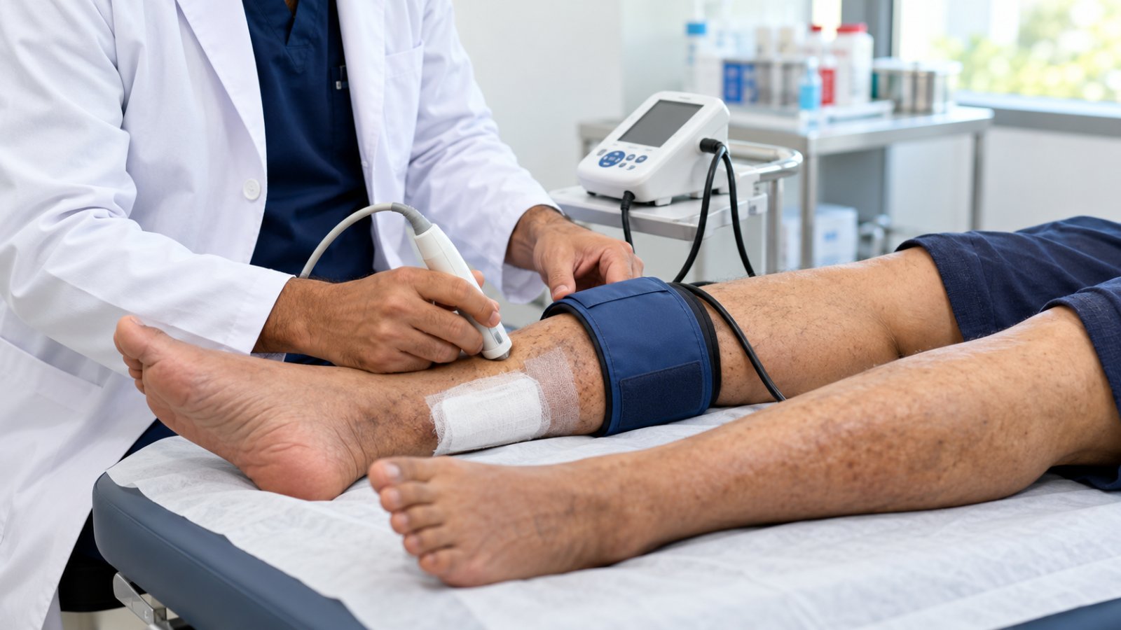
An ankle-brachial pressure index compares systolic blood pressure measured at the ankle with pressure measured in the arm.
It helps identify reduced arterial blood flow.
However, ABPI may be falsely elevated when arteries are stiff or calcified, particularly in people with diabetes or chronic kidney disease.
In such cases, toe pressures, toe-brachial index or other vascular investigations may be required.
Step 3: Investigate Venous Disease
Venous duplex ultrasound evaluates blood flow through the leg veins.
It can identify:
-
Venous reflux
-
Venous obstruction
-
Previous thrombosis-related changes
-
Other abnormalities affecting venous drainage
These findings help determine whether compression, venous intervention or other treatment is appropriate.
Patients can find more information about vascular assessment.
How Do Venous, Arterial, Diabetic and Vasculitic Ulcers Differ?
The underlying cause determines the most appropriate treatment.
A chronic leg ulcer should not automatically be labelled venous or diabetic without assessment.
1. Venous Leg Ulcers
Venous ulcers develop when the veins cannot return blood efficiently toward the heart.
This increases pressure within the venous circulation.
Typical features include:
-
Ulcers around the lower leg or ankle
-
Leg swelling
-
Brown skin pigmentation
-
Varicose veins
-
Skin thickening
-
Irregular or shallow wound edges
-
Discomfort that may improve with leg elevation
Compression therapy is a major component of treatment when arterial circulation is adequate.
Patients can read more about venous leg ulcers.
2. Arterial Leg Ulcers
Arterial ulcers occur when reduced blood flow limits the delivery of oxygen and nutrients to tissues.
Possible features include:
-
Painful ulcers on the foot, toes or pressure points
-
Cool skin
-
Weak or absent foot pulses
-
Shiny or thin skin
-
Pain that worsens with elevation
-
Tissue loss or blackened areas
Significant arterial disease requires vascular assessment.
Standard high-strength compression may be unsafe when arterial circulation is severely reduced.
3. Diabetic Ulcers
Diabetes-related ulcers commonly develop because of peripheral neuropathy, pressure and sometimes peripheral arterial disease.
Typical findings may include:
-
Reduced foot sensation
-
Repeated unnoticed injuries
-
Callus formation
-
Ulcers beneath pressure-bearing areas
-
Foot deformity
-
Delayed wound healing
Treatment may require pressure offloading, infection management, glucose control and circulation assessment.
Not every ulcer in a person with diabetes is caused by diabetes alone.
4. Vasculitic Ulcers
Vasculitis involves inflammation of blood vessels.
Inflammation can damage vessel walls and reduce blood supply to surrounding tissue.
Possible signs include:
-
Painful skin ulcers
-
Purple or reddish skin lesions
-
Palpable purpura
-
Tissue breakdown
-
Multiple lesions
-
Systemic symptoms such as fever or joint pain
Appropriate vasculitis ulcer treatment depends on identifying the underlying inflammatory condition.
Some patients require biopsy, blood tests and specialist-directed medication.
More information is available on autoimmune and vasculitis ulcers.
Comparison of Common Leg Ulcer Types
| Ulcer Type | Common Clues | Important Investigation | Main Treatment Principle |
|---|---|---|---|
| Venous | Swelling, pigmentation, lower-leg ulcer | ABPI and venous duplex | Compression when safe; address venous disease |
| Arterial | Pain, cool skin, reduced pulses | ABPI, toe pressures and vascular imaging | Restore or improve circulation when indicated |
| Diabetic | Neuropathy, callus, pressure-related wound | Sensory and vascular assessment | Offloading, wound care and metabolic management |
| Vasculitic | Severe pain, purpura, atypical lesions | Selected blood tests and biopsy | Treat underlying inflammation |
Mixed-cause ulcers are also possible.
For example, a patient may have both venous insufficiency and peripheral arterial disease.
Which Clues Point to Vasculitis, Pyoderma or Cancer?
Some ulcers do not follow the typical appearance or healing pattern of common venous or diabetic wounds.
These are often described as atypical ulcers.
Recognising unusual features is important because standard wound treatment may be insufficient or inappropriate.
Atypical Ulcer Red Flags
Seek specialist assessment when an ulcer has:
-
Rapid enlargement
-
Severe pain disproportionate to its appearance
-
Purple or undermined borders
-
Palpable purpura
-
Unusual location
-
Raised or rolled wound edges
-
Excessive or abnormal granulation tissue
-
Unexplained bleeding
-
Recurrent breakdown at the same site
-
Unexpected deterioration despite appropriate care
-
Associated fever, joint pain or unexplained weight loss
These findings do not establish a specific diagnosis, but they indicate the need for further investigation.
Could It Be Pyoderma Gangrenosum?
Pyoderma gangrenosum is an inflammatory skin condition that can cause rapidly progressive, painful ulcers.
A characteristic feature may be a violaceous or undermined wound border.
Some patients experience worsening after trauma, a phenomenon called pathergy.
Because of this, aggressive surgical debridement before the condition is recognised may worsen the ulcer.
A dermatologist or other appropriate specialist should evaluate suspected cases.
Could a Chronic Ulcer Be Skin Cancer?
Yes.
Certain skin cancers can present as persistent ulcers or develop in longstanding wounds.
Warning signs include:
-
Raised or rolled edges
-
Unusual tissue growth
-
Persistent bleeding
-
Progressive enlargement
-
Failure to heal despite appropriate treatment
A biopsy may be required to exclude malignancy.
Patients with unexplained wounds may also benefit from information about chronic ulcers of unknown cause.
When Is a Chronic Leg Ulcer Biopsy Needed?
A biopsy involves removing a small sample of tissue for laboratory examination.
It can help identify inflammatory disease, vasculitis, malignancy and other uncommon causes.
When Should a Biopsy Be Considered?
A biopsy may be indicated when:
-
The ulcer has an unusual appearance.
-
Cancer is suspected.
-
Vasculitis is suspected.
-
The wound deteriorates unexpectedly.
-
The diagnosis remains unclear.
-
The ulcer does not improve after an adequate period of correctly directed treatment.
What Is the 12-Week Biopsy Rule?
An ulcer that has not shown meaningful improvement after approximately 12 weeks of appropriate treatment should undergo diagnostic reassessment, including consideration of biopsy.
However, 12 weeks is not a mandatory waiting period.
If an ulcer has suspicious features, biopsy or specialist referral may be needed much earlier.
The decision also depends on wound duration, prior treatment, circulation and clinical appearance.
Does Every Chronic Ulcer Need a Biopsy?
No.
A typical venous ulcer that responds to appropriate treatment may not require biopsy.
The question of when to biopsy a leg ulcer depends on the clinical findings and healing response.
If vasculitis is suspected, biopsy technique and selection of the tissue sample are especially important.
The specialist may sample an active lesion and request additional testing.
Why Must Arterial Disease Be Ruled Out Before Compression?
Compression therapy is widely used for venous leg ulcers.
It applies controlled external pressure to help improve venous return and reduce swelling.
However, compression must be prescribed carefully.
What Happens If Arterial Circulation Is Poor?
When arterial blood supply is significantly reduced, strong compression can further compromise tissue perfusion.
This may increase pain or worsen tissue damage.
Therefore, clinicians should assess arterial circulation before prescribing therapeutic compression.
Understanding ABPI Results
| ABPI Result | General Interpretation | Compression Consideration |
|---|---|---|
| Approximately 0.8–1.3 | Often compatible with adequate arterial circulation | Standard compression may be appropriate after assessment |
| Approximately 0.5–0.8 | Suggests mixed arterial and venous disease | Modified compression may be considered under specialist supervision |
| Below approximately 0.5 | Suggests severe arterial disease | Avoid routine compression and seek urgent vascular assessment |
| Above approximately 1.3 | May indicate incompressible, calcified arteries | Additional tests such as toe pressures may be needed |
These are general thresholds, not standalone prescribing instructions.
Actual compression decisions also depend on absolute ankle pressure, toe pressure, symptoms and the patient's overall vascular status.
Can Patients With Mixed Ulcers Receive Compression?
Sometimes.
Patients with both venous and arterial disease may benefit from carefully selected, reduced-pressure compression when arterial perfusion is sufficient.
However, this requires specialist assessment.
Patients should not independently apply strong compression bandages to an ulcer without appropriate circulation testing.
Non Healing Leg Ulcer Treatment in Vizianagaram: When to Seek Specialist Care
Patients with a wound that remains open for several weeks should seek a structured wound and vascular assessment.
For those searching for non healing leg ulcer treatment in vizianagaram, the priority is identifying whether the ulcer is venous, arterial, diabetic, inflammatory or caused by another condition.
At Elegance Diabetic Foot & Ulcer Clinic (EDFC), Dr. Ashutosh Shah is associated with assessment and management of complex lower-limb wounds. Evaluation may involve wound examination, circulation testing and appropriate specialist investigations based on the suspected cause.
Patients should confirm consultation locations and available investigations directly with the clinic.
What Happens During the First Assessment?
A clinician may:
-
Review the wound history
-
Examine both legs and feet
-
Assess pain and swelling
-
Check peripheral pulses
-
Evaluate sensation
-
Measure the wound
-
Review diabetes and other medical conditions
-
Request appropriate investigations
-
Develop a cause-specific treatment plan
The aim is to establish a diagnosis rather than repeatedly changing dressings without addressing the underlying problem.
Why Is Regular Wound Measurement Important?
Documenting ulcer dimensions helps determine whether treatment is working.
Measurements may include:
-
Length
-
Width
-
Depth
-
Wound area
-
Tissue appearance
-
Exudate
-
Surrounding skin condition
Follow-up measurements may be taken approximately every two weeks, or more frequently when clinically indicated.
If the wound is not improving, the team should reassess the diagnosis, circulation, pressure exposure, infection status and treatment adherence.
What Treatment Helps a Chronic Leg Ulcer Heal?
Effective chronic leg ulcer management depends on the diagnosis.
Venous Ulcers
Treatment may involve:
-
Compression after arterial assessment
-
Appropriate dressings
-
Leg elevation
-
Exercise and calf-muscle activity
-
Venous intervention when indicated
Arterial Ulcers
Treatment may involve:
-
Vascular imaging
-
Medical management of cardiovascular risk
-
Revascularisation when appropriate
-
Protective wound care
-
Avoidance of harmful pressure
Diabetic Ulcers
Treatment may involve:
-
Pressure offloading
-
Glucose management
-
Infection assessment
-
Debridement when appropriate
-
Vascular evaluation
-
Protective footwear
Vasculitic and Inflammatory Ulcers
Treatment may require:
-
Dermatology or rheumatology assessment
-
Biopsy when indicated
-
Evaluation for systemic disease
-
Specialist-directed anti-inflammatory or immunosuppressive treatment
-
Appropriate wound care
No single dressing, cream or procedure is suitable for every ulcer.
Conclusion
A chronic leg ulcer that remains open for more than six weeks should not be managed with dressing changes alone.
The underlying cause must be identified through clinical examination and appropriate investigations.
ABPI helps assess arterial circulation, venous duplex identifies venous abnormalities, and selected blood tests evaluate diabetes, kidney function and inflammation.
Unusual ulcers or wounds that fail to improve may require biopsy.
Most importantly, compression therapy should not be prescribed without considering arterial circulation.
At Elegance Diabetic Foot & Ulcer Clinic, Dr. Ashutosh Shah is associated with evaluation and treatment planning for complex lower-limb wounds.
Early diagnosis, appropriate treatment and regular wound monitoring can help reduce complications and improve healing outcomes.
quiz Frequently Asked Questions
This article is general education, not a diagnosis. If you have a diabetic foot wound, please have it assessed in person. Send a photo on WhatsApp or book a consultation.
About the Author

Dr. Ashutosh Shah, Plastic, Reconstructive & Diabetic Foot Surgeon, Elegance Diabetic Foot & Ulcer Clinic, Surat
Dr. Ashutosh Shah is a board certified plastic and reconstructive surgeon with over 22 years of experience and more than 10,000 limbs and feet saved. He founded Elegance Diabetic Foot & Ulcer Clinic, India's first chain of clinics dedicated to foot care, with centres in Surat and OPD partners across South Gujarat. He is known for a limb first approach, safe wound care and ethical, natural results.