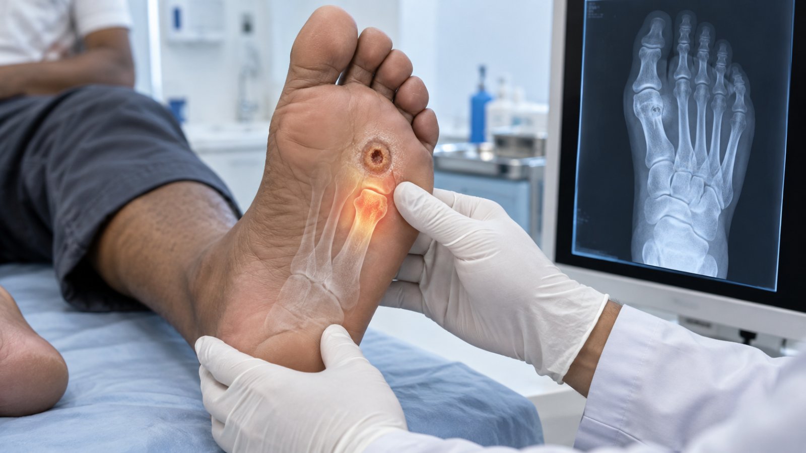
If a sterile probe touches bone through the ulcer, bone infection is likely. X-rays stay normal for two to three weeks because bone must lose about half its mineral before changes show. MRI detects it earlier, and bone biopsy confirms which organism is present.
At Elegance Diabetic Foot & Ulcer Clinic (EDFC), Dr. Ashutosh Shah and the diabetic foot care team assess deep or non-healing ulcers for possible bone infection diabetic foot complications. The wound depth, probe-to-bone findings, clinical signs, imaging and laboratory results are considered together rather than relying on a normal early X-ray alone.
What Is the Probe-to-Bone Test and How Reliable Is It?
The probe-to-bone test is a simple clinical test used when a diabetic foot ulcer is deep enough for underlying bone involvement to be a concern.
During the test, a clinician gently introduces a sterile blunt metal probe through the ulcer. If hard or gritty bone can be felt at the base of the wound, the test is considered positive.
A positive result raises suspicion of osteomyelitis, particularly when the ulcer is deep, chronic or already shows signs of infection.
However, the probe-to-bone test should not be interpreted on its own. Its significance depends on the wound and the patient's overall clinical picture. A positive test increases concern for bone infection diabetic foot, while a negative test does not automatically exclude infection in every patient.
The result may therefore need to be combined with imaging, blood tests and, in selected cases, bone sampling.
Read more about Osteomyelitis in the diabetic foot.
Why Does an Early X-Ray Often Look Normal?
A plain X-ray is commonly one of the first imaging tests requested when bone infection is suspected.
The difficulty is timing.
According to the diagnostic framework in this brief, X-ray changes may remain absent for the first two to three weeks because substantial mineral loss must occur before the infection becomes clearly visible on a plain radiograph.
This means a patient may have clinically important bone infection even though an early X-ray appears normal.
As infection progresses, an X-ray may eventually show changes involving the bone. It can also provide useful information about deformity, fractures, foreign material or other structural abnormalities.
But when there is strong clinical suspicion of bone infection diabetic foot, a normal early X-ray should not automatically end the investigation.
Further Imaging for the diabetic foot may be required when the examination and initial X-ray do not provide a clear answer.
When Is an MRI Needed?
MRI can detect changes associated with bone and soft-tissue infection earlier and in greater anatomical detail than a plain X-ray.
It becomes particularly useful when there is continuing suspicion of osteomyelitis but the diagnosis remains uncertain after the clinical examination, probe-to-bone test and initial X-rays.
MRI can help the treating team assess:
- which bone may be involved
- the extent of abnormal bone changes
- surrounding soft-tissue infection
- deeper collections
- the relationship between the ulcer and underlying structures
An MRI is not necessarily required for every diabetic foot ulcer.
A superficial ulcer without findings suggesting deeper infection may not need advanced imaging. The decision should be based on ulcer depth, clinical findings and the likelihood that identifying deeper infection will change treatment.
For suspected bone infection diabetic foot, MRI is particularly valuable when earlier steps leave uncertainty about whether infection has reached the bone or how far it extends.
What Does a Bone Biopsy Add?
Imaging can suggest osteomyelitis, but it does not necessarily identify the organism causing the infection.
A bone biopsy can provide more specific information.
A sample of bone is obtained and sent for microbiological assessment. This can help determine which organism is present and guide antibiotic selection. Depending on the clinical situation, the bone may also be examined for changes consistent with infection.
This distinction matters because bacteria found on the surface of a chronic ulcer are not necessarily the same organisms causing infection deeper in the bone.
Bone biopsy therefore has a different role from an MRI. MRI helps identify the location and extent of suspected infection, while bone sampling can help establish what is actually present within the bone.
Other Laboratory tests may also contribute to the assessment of infection and the patient's overall condition.
Diagnostic Ladder for Suspected Bone Infection
| Diagnostic step | What it confirms or suggests | What it can miss | When to move to the next step |
|---|---|---|---|
| Clinical signs | Identifies a deep or infected ulcer and raises suspicion of deeper involvement | Bone infection may exist without dramatic surface signs | Move on when the ulcer is deep, chronic or suspicious |
| Probe to bone | Bone felt through the ulcer raises suspicion of osteomyelitis | A negative test does not exclude every case | Proceed when suspicion remains or the test is positive |
| Plain X-ray | May show established bone changes and other structural abnormalities | Early infection may look normal | Consider MRI when suspicion remains despite an inconclusive X-ray |
| MRI | Helps define suspected bone and soft-tissue involvement | Does not by itself identify the causative organism | Consider bone sampling when microbiological confirmation will change treatment |
| Bone biopsy | Helps identify organisms present in the bone and supports definitive diagnosis | Sampling may not be necessary in every patient | Use results with the clinical picture to guide treatment |
| Spreading infection signs | Fever, rigors, red streaks or rising swelling may indicate infection extending beyond the local area | — | Go to hospital the same day |
How Does Bone Involvement Change Antibiotic Duration and Surgery?
A superficial soft-tissue infection and osteomyelitis are not treated as if they are the same problem.
Once infection reaches bone, treatment may become longer and more complex. Antibiotic choice and duration depend on factors such as the extent of infection, organisms identified, whether infected bone is removed and the patient's overall condition.
Some patients can be managed without removing major parts of the foot. Others may require surgical removal of infected or dead bone when infection cannot be adequately controlled by medical treatment alone.
Surgery may also be considered when there is:
- dead or severely infected bone
- an abscess or deep collection
- progressive tissue destruction
- infection that does not respond adequately to treatment
- mechanical or structural problems preventing wound healing
A diagnosis of bone infection diabetic foot does not automatically mean amputation. The treatment plan depends on the amount of bone involved, circulation, soft-tissue condition, infection severity and whether a functional foot can be preserved.
Appropriate Infection management may therefore involve antibiotics, wound treatment, pressure relief, surgery or a combination of these approaches.
Which Signs Mean the Infection Is Spreading Beyond the Bone Right Now?
Osteomyelitis can sometimes remain relatively localised, but a diabetic foot infection can also spread into surrounding tissues or become a systemic illness.
This is when waiting for a routine appointment can become dangerous.
Seek same-day hospital assessment if there is:
- fever
- rigors or shaking chills
- rapidly increasing swelling
- redness spreading across the foot or up the leg
- red streaks extending away from the infected area
- rapidly increasing discharge
- foul smell with worsening wound appearance
- sudden tissue discolouration
- increasing weakness, confusion or systemic illness
A person with neuropathy may not experience severe pain even when infection is progressing. Lack of pain should therefore never be used as proof that a deep diabetic foot infection is safe.
When bone infection diabetic foot is accompanied by fever, rigors, spreading redness or rapidly increasing swelling, the priority changes from routine investigation to urgent assessment and infection control.
Conclusion: A Normal Early X-Ray Does Not End the Investigation
Suspected bone infection diabetic foot needs a step-by-step assessment. A deep ulcer, positive probe-to-bone test or other concerning findings may raise suspicion even when an early X-ray looks normal.
The diagnostic pathway can progress from clinical examination and probe-to-bone testing to plain X-ray, MRI and, when required, bone biopsy. Each step answers a different question: whether bone involvement is likely, where the infection has spread and which organism may be responsible.
Most importantly, fever, rigors, red streaks, rapidly increasing swelling or other signs of spreading infection should not wait for routine imaging or the next clinic visit. These changes require same-day medical assessment.
quiz Frequently Asked Questions
This article is general education, not a diagnosis. If you have a diabetic foot wound, please have it assessed in person. Send a photo on WhatsApp or book a consultation.
About the Author

Dr. Ashutosh Shah, Plastic, Reconstructive & Diabetic Foot Surgeon, Elegance Diabetic Foot & Ulcer Clinic, Surat
Dr. Ashutosh Shah is a board certified plastic and reconstructive surgeon with over 22 years of experience and more than 10,000 limbs and feet saved. He founded Elegance Diabetic Foot & Ulcer Clinic, India's first chain of clinics dedicated to foot care, with centres in Surat and OPD partners across South Gujarat. He is known for a limb first approach, safe wound care and ethical, natural results.